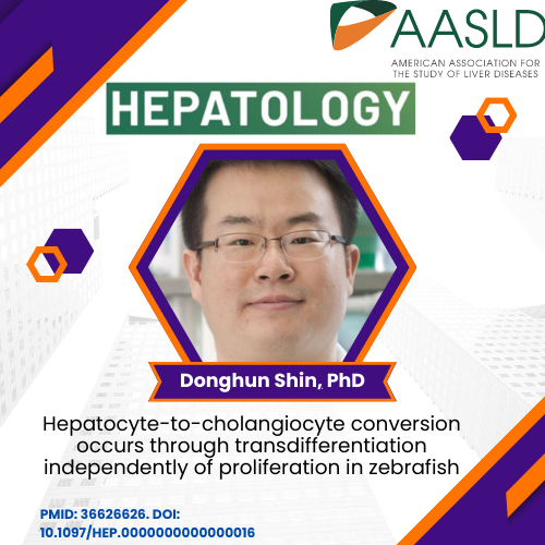
Donghun Shin, PhD., Associate Professor in the department of Developmental Biology has done fundamental work in understanding liver development and cellular differentiation. He recently published an article in Hepatology entitled, “Hepatocyte-to-cholangiocyte conversion occurs through transdifferentiation independently of proliferation in zebrafish.” In addition he co-authored a review in Seminars in Liver Disease entitled, “Update on Hepatobiliary Plasticity“.
Abstract
Background and aims: Injury to biliary epithelial cells (BECs) lining the hepatic bile ducts leads to cholestatic liver diseases. Upon severe biliary damage, hepatocytes can convert to BECs, thereby contributing to liver recovery. Given a potential of augmenting this hepatocyte-to-BEC conversion as a therapeutic option for cholestatic liver diseases, it will be important to thoroughly understand the cellular and molecular mechanisms of the conversion process.
Approach and results: Towards this aim, we have established a zebrafish model for hepatocyte-to-BEC conversion by employing Tg(fabp10a:CFP-NTR) zebrafish with a temporal inhibition of Notch signaling during regeneration. Cre/loxP-mediated permanent and H2B-mCherry-mediated short-term lineage tracing revealed that in the model, all BECs originate from hepatocytes. During the conversion, BEC markers are sequentially induced in the order of Sox9b, Yap/Taz, Notch activity/ epcam , and Alcama/ krt18 ; the expression of the hepatocyte marker Bhmt disappears between the Sox9b and Yap/Taz induction. Importantly, live time-lapse imaging unambiguously revealed transdifferentiation of hepatocytes into BECs: hepatocytes convert to BECs without transitioning through a proliferative intermediate state. In addition, using compounds and transgenic and mutant lines that modulate Notch and Yap signaling, we found that both Notch and Yap signaling are required for the conversion even in Notch- and Yap-overactivating settings.
Conclusions: Hepatocyte-to-BEC conversion occurs through transdifferentiation independently of proliferation, and Notch and Yap signaling control the process in parallel with a mutually positive interaction. The new zebrafish model will further contribute to a thorough understanding of the mechanisms of the conversion process.
Update on Hepatobiliary Plasticity
- Kim M, Rizvi F, Shin D, Gouon-Evans V. Update on Hepatobiliary Plasticity. Semin Liver Dis. 2023 Feb 10. doi: 10.1055/s-0042-1760306. Epub ahead of print. PMID: 36764306.https://www.thieme-connect.com/products/ejournals/html/10.1055/s-0042-1760306
Abstract
The liver field has been debating for decades the contribution of the plasticity of the two epithelial compartments in the liver, hepatocytes and biliary epithelial cells (BECs), to derive each other as a repair mechanism. The hepatobiliary plasticity has been first observed in diseased human livers by the presence of biphenotypic cells expressing hepatocyte and BEC markers within bile ducts and regenerative nodules or budding from strings of proliferative BECs in septa. These observations are not surprising as hepatocytes and BECs derive from a common fetal progenitor, the hepatoblast, and, as such, they are expected to compensate for each other’s loss in adults. To investigate the cell origin of regenerated cell compartments and associated molecular mechanisms, numerous murine and zebrafish models with ability to trace cell fates have been extensively developed. This short review summarizes the clinical and preclinical studies illustrating the hepatobiliary plasticity and its potential therapeutic application.
Graphical Abstract:


